From Lab Bench to Sofa
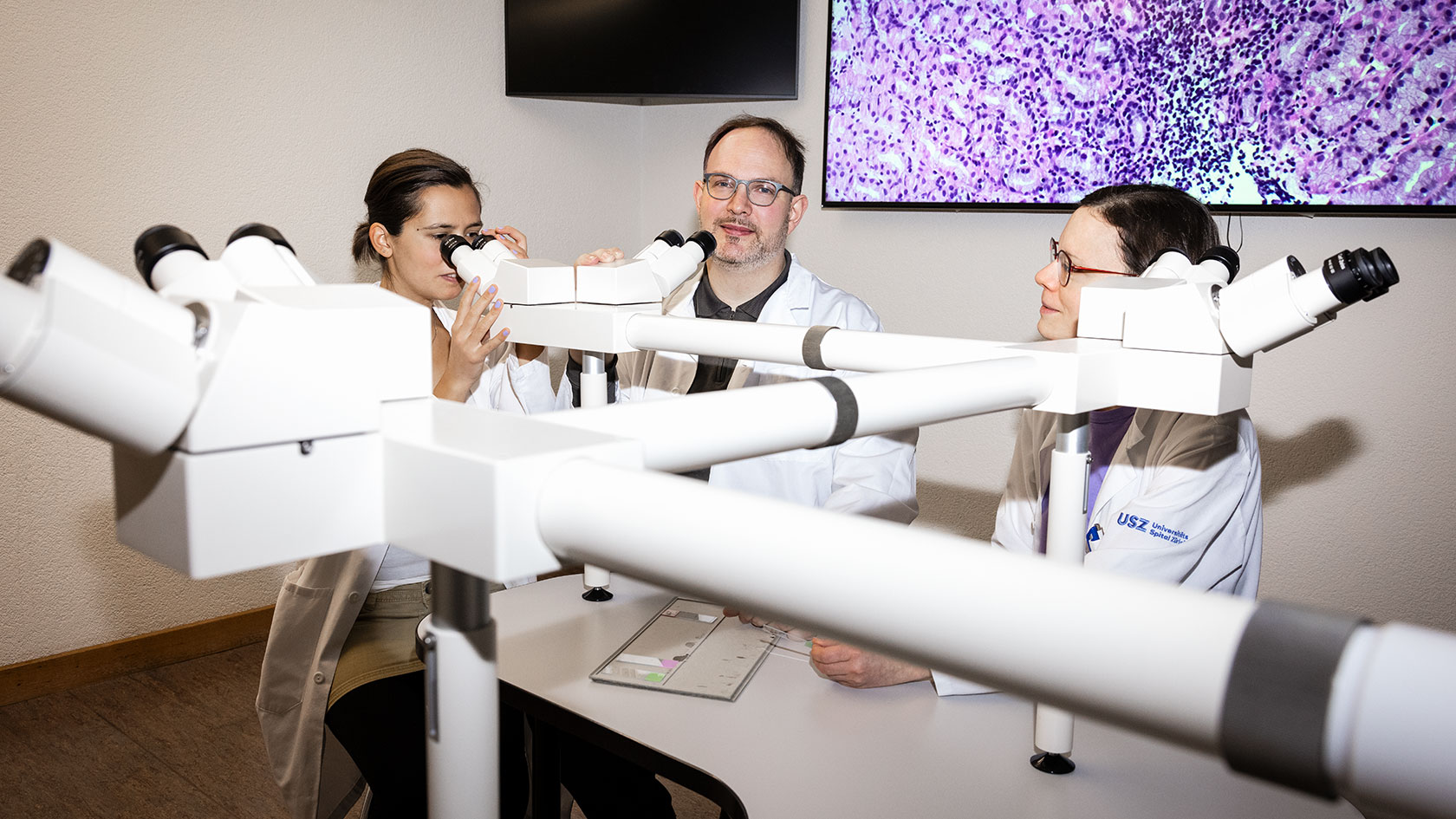
Medical students in white lab coats sit in rows, each peering into a microscope: an image that seems like a throwback to histopathology courses of the early 20th century. Nowadays, students are more likely to be sitting comfortably on the sofa at home and acquiring their subject knowledge online. Soon they will even be able to follow microscope examinations from the comfort of their homes.
Together with the Institute of Molecular and Cellular Anatomy at Ulm University, Michael Reinehr, clinical instructor at UZH and pathologist at the UniversityHospital Zurich, has developed the MyMi.mobile 2.0 platform with which students can examine a variety of specially colored tissue samples. The app contains high-resolution scans of specimens around which students can freely navigate and zoom in and out of, as if using a virtual microscope.
“Until now, students had usually only seen the samples once before sitting an exam,” says Reinehr. “The app gives students unlimited access to the specimen collection, meaning they can prepare for the course and follow up afterward on their mobile or laptop – they could even use their journey home to learn more!”
From organ to cell
Reinehr noticed when teaching that histopathology posed quite a challenge for students new to the subject. He therefore decided to try to create a tool to help them make the most of their self-study time. Histopathology is about diagnosing diseases based on observable changes at the cellular level.
To make a diagnosis, surgically removed tissue is cut into wafer-thin strips, dyed and examined under the microscope. “It’s easy to understand how a heart works if you have the whole organ in front of you,” says Reinehr. “But if you are only looking at a small piece and zooming in to the cellular level, it becomes abstract and difficult to comprehend.”
Understanding the connection between cell structure and organ function or dysfunction is the overarching learning objective of histopathology courses in the third, fourth and sixth years of medical school. In his teaching, Reinehr is therefore always jumping back and forth between the macro and micro levels. “In the case of cirrhosis, where we have a liver riddled with fibrous connective tissue, we show students the whole organ first,” he explains.
“They see the nodules on the cross-section and notice that they’re turning green because of bile stasis.” This overview makes it easier for the students to later classify the corresponding specimen and to understand why they can see connective tissue fibers and bile droplets under the microscope. “Such connections are difficult to comprehend if students only see the specimens once without being able to take the time – for example with MyMi.mobile 2.0 – to get to grips with what they’re observing,” says Reinehr.
Interactive classes despite high student numbers
The idea for the virtual microscope came from Stefan Britsch, professor of anatomy at Ulm University, under whom Reinehr previously worked. Britsch developed MyMi’s predecessor, MyMicroscope, over a decade ago, for which he scanned histology samples for the first time. “At the time it was groundbreaking: it was the first time the internet had been used for something like this,” says Reinehr.
The new version of the program is run through the powerful computing center of Ulm University and also has an improved viewer. This means many students can access the software simultaneously without slowing down the program. In the future, it will thus also be possible to conduct exams – for example, the pathology exam required for admission to the state board examination for medicine – via the platform.
Thanks to a grant from the UZH Teaching Fund, Reinehr was able to acquire an unlimited license to use the platform. Reinehr then spent 18 months collecting interesting diseased tissue samples from routine diagnostic processes, before scanning them and uploading them to the app for students to examine.
“This means students see realistic cases rather than textbook-perfect ones,” he explains. He sifted through the storage stacks to find 200 interesting specimens suitable for teaching pathology. These had to be perfectly cut and then scanned. “I underestimated the amount of work it would take,” he says. The scans are currently waiting to be processed so that they will be available to the students in high-resolution quality. “Then, during the online histopathology course, students will be able to follow the microscope investigation from home as if they were doing it themselves in the lab.”
Individual feedback thanks to AI
The MyMI.mobile 2.0 platform also has other uses outside of class time. Its main purpose is to enable students to learn independently. For each specimen, they can look at an overlay with notes and arrows marking areas of interest. They can then read more about these cell structures via a drop-down menu. “We can link textbook chapters or interesting papers here too,” says Reinehr.
Being able to view the tissue samples on a computer or tablet also makes it easier for students to discuss them in study groups. In addition, the platform has practice exercises in which students have to search for certain structures or make a diagnosis. The program gives them immediate feedback and shows them what percentage they answered correctly.
The feedback function is currently being expanded by the German Research Centre for Artificial Intelligence. They are working on an algorithm that will monitor students’ learning progress and provide them with individualized feedback. “The algorithm will track learners’ methods of searching for structures, and their progress – with the users’ consent of course,” says Reinehr. These data will also help instructors improve their classroom teaching. In the future, a virtual tutor function may be added to the platform to give students additional support.
Exploring cell structures with an audio guide
Four universities from Germany are already using the platform for their anatomical specimen collection. The University of Zurich is the first partner to upload histopathology samples. Students also have access to the collections of other universities, allowing them to practice with other specimens too.
With just a few mouse clicks, they can skip from a histopathological view of liver cirrhosis to the tissue sample of a healthy liver and compare them with each other. This feature enables students to link histopathology to their prior knowledge of anatomy, thereby promoting successful learning.
“The platform is based on a principle of sharing: each university benefits from the information uploaded by the others,” says Reinehr. Next, he plans to upload macro images of organs so that students can directly compare the complete organ with tissue samples.
Reinehr is full of ideas about how to expand the platform in the future. Radiology scans could be uploaded, for example. It would then be possible to look at the CT cross-section image of a diseased liver alongside the histopathology sample plus a healthy liver for comparison. “Such a step would really facilitate cross-curricular teaching,” believes Reinehr. He also plans to record histopathology podcasts in which he will analyze specimens and explain his pathologist’s reading of them. The students will be looking at the microscope view as they listen, so that the podcast provides a kind of audio guide to the specimen.
Paradigm shift in pathology
Digitalization of specimens and diagnosis via computer screen has become common practice for students, and now clinical pathology is starting to catch up. In the future, specimens at USZ will be directly scanned and made available to the pathologists on their computers. “First, however, the logistics of dealing with such large volumes of data need to be solved and a functioning workflow established,” says Reinehr.
Digitalization brings many advantages for clinical pathologists. One benefit concerns storage space, since currently all samples must be physically stored in wax blocks and in the form of glass slides for 10 to 15 years.
Another is that researchers no longer need to travel to view samples, and it is easier to discuss a tricky specimen with a specialist in another part of the world. Digital pathology is fundamentally transforming the field – for example, pattern recognition by artificial intelligence could also be used to support diagnostics in the future.
Michael Reinehr’s day would then look quite different. Instead of spending the day peering into a microscope in the lab, he could make diagnoses at home from behind his computer.