Watching the Brain Learn
What could be better than finding your own idea make its way into the world? This is exactly what Fritjof Helmchen and his assistant Nikita Vladimirov are currently experiencing. They’re working on the development of a special microscope at the Brain Research Institute at UZH. Their “mesoSPIM” makes it possible to create a three-dimensional image of brain tissue in just a few minutes – this may involve entire mouse brains or samples of tissue from human brains. They are constantly improving the microscope. They make the instructions explaining how to construct the latest version freely accessible on the internet. Research institutions all over the world are making use of these instructions, with more than thirty replica versions already in existence.
This is no wonder: “Without imaging processes, brain research is impossible,” says Esther Stoeckli, professor in the Department of Molecular Life Sciences at UZH. Together with Helmchen, she leads the University Research Priority Program (URPP) “Adaptive Brain Circuits in Development and Learning” (see box). There are two questions at the heart of this network comprising a total of 23 research groups at UZH: what happens in the brain when it develops, and how does it change when we learn? What are the causes of learning disorders and how can they be treated better in the future?
Novel microscopes
Imaging techniques are key to the researchers. “Modern brain research began with microscopy around 150 years ago. And today, we’re embarking on a new era, mainly thanks to improved imaging techniques, that will give us a better and better understanding of the brain,” says Helmchen. Newly developed techniques for staining individual cells enabled specific parts of the brain to be visualized using light microscopy for the first time in the 19th century. Back then, people examined flat slices of dissected brains.
A lot has happened since then. For example, the very-high-resolution electron microscope was created. In addition, novel microscopes were developed in order to use laser light to scan tissue layer by layer, which produces 3D views instead of just being able to look at specimens in the form of slices.
Biology and AI inspire each other
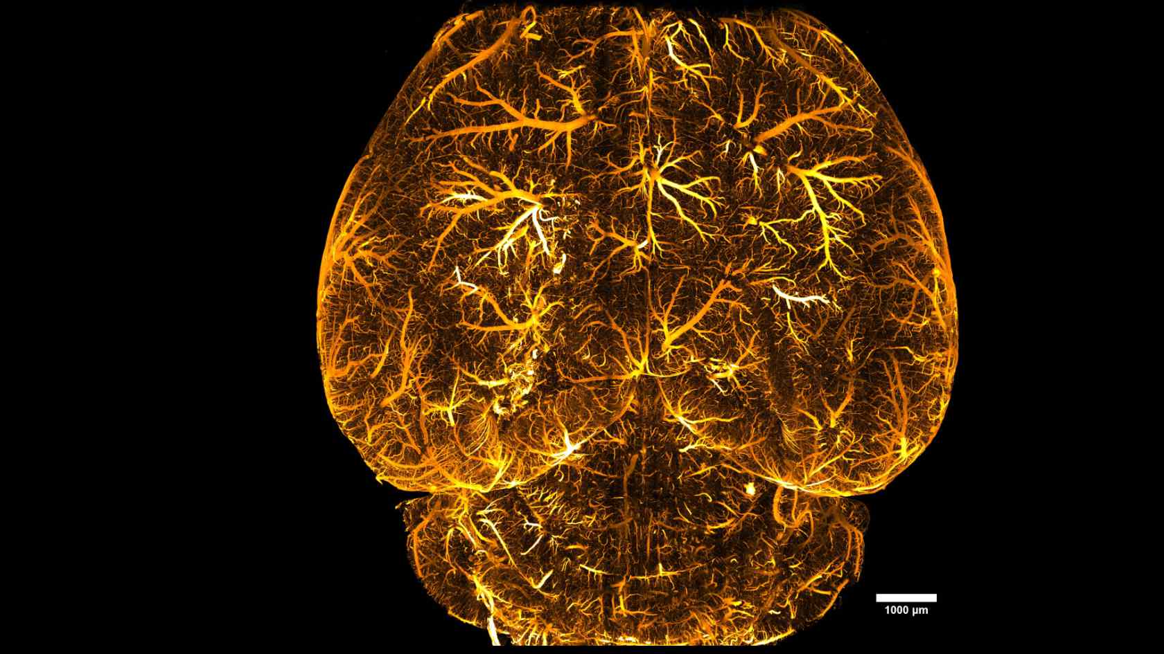
Nowadays, a large number of different microscopes are used in brain research. They include multi-photon fluorescence microscopes and light sheet microscopes – the latter category includes the mesoSPIM from the Helmchen research group. Both types of microscopes make it possible to view the anatomy of the brains of laboratory animals in high resolution down to the level of individual nerve cells and synapses.
Multi-photon microscopy also makes it possible to measure the activity patterns in the networks of nerve cells. When it comes to watching the human brain at work, functional magnetic resonance imaging (fMRI) is also used. This technique measures changes in the blood flow in the brain, allowing conclusions to be drawn about which areas of the brain are particularly active during certain tasks.

Modern brain research began with microscopy around 150 years ago. Today, we’re embarking on a new era, thanks to improved imaging techniques, that will give us a better and better understanding of the brain.
Novel microscopes were not the only key factor behind the growing importance of imaging in brain research. There were also other developments: for example, it became possible to chemically modify brain specimens to make them transparent. This means that even larger blocks of tissue can be visualized without needing to cut them open. And last but not least, advances in computer science now make it possible to process the ever-larger amounts of data that the imaging produces.
Today, we’re also seeing biology and AI inspiring each other: new findings from the biological brain are making it possible to adapt machine learning methods to reflect them and simulate functions of the brain. Conversely, AI enables the construction of neural networks, such as those you might find in the brain, if you can search specifically for them.
“Today we can do almost anything: we can look deeper into the brain, observe processes instead of just momentary snapshots and also look at smaller structures in ever higher resolution,” says Stoeckli. In addition, the processes are faster because complex tissue preparations, of the kind needed for electron microscopy, are often no longer required with modern microscopes. However, many methods can only be used on animals. For example, ethical reasons dictate that it’s not possible to stain cells or perform genetic modifications in humans. But imaging on its own is not capable of explaining the advances in brain research. Genetic and molecular biological approaches along with methods such as electrophysiology, which records electrical impulses in the brain, have also become increasingly important.
How does the brain work? Insights into the research of Prof. Dr. Fritjof Helmchen in the video.
Production: MELS, University of Zurich
Deeper, sharper, faster
The group led by Helmchen is one of the world’s leading groups researching the evolution of imaging technologies. It’s creating new types of lenses and detectors for microscopes and programming the relevant software needed for data processing. But above all, Helmchen, his mesoSPIM specialist Nikita Vladimirov and the team are constantly looking for further ways to keep developing their microscope.
“Each advance in technology also allows us to gain new insights into the brain,” says Esther Stoeckli. For example, we now know about lots of different types of nerve cells in the brain. It has also been possible to reconstruct the brain of a fly as a complete network of all neurons with all their connections – known as the connectome. Helmchen’s assessment is that this should soon also be possible in the model organism of the zebrafish larva and, within five to ten years, in mice: “But in humans, this is still a long way off.”
Esther Stoeckli and Fritjof Helmchen focus on different issues. Stoeckli mainly investigates the development of the brain from the embryo to the adult animal or human. She’s particularly interested in how the axons – the extensions of nerve cells – find their way to their target cells, to which they are supposed to transmit information. This process is crucial for the formation of neural networks. Helmchen primarily conducts research into the processes in the already developed brain. Among other aspects, his group investigates how the signal propagation, which means the temporal sequence of the activity of nerve cells in different regions of the brain, changes during learning processes.
The challenge is to display ever larger parts of the brain in ever higher resolution. Larger specimens are fascinating because, in many processes, different parts of the brain are active at the same time or in coordination and should be considered as such. The intention is to observe the processes down to the level of the individual synapses, which are only about a thousandth of a millimeter in size. The researchers sometimes take unusual paths to achieve both these objectives wherever possible. Instead of just focusing on improving the resolution of the microscopes, they’ve recently also started enlarging the specimens using special biotechnological methods. This process involves “inflating” the cells.
A model of brain function
The goal of the URPP is still to understand the healthy brain so that learning disorders can then also be better understood. Here, too, many questions remain unanswered. Thanks to new findings, the vision is to develop more effective therapies for treating learning disorders and new learning methods in the future. “To achieve this goal, we need to be able to create an overarching theoretical model of brain function,” says Helmchen. This is an attempt to represent our thinking organ in mathematical formulas.
Helmchen and Stoeckli agree that it will be many years before this happens. Just watching the brain passively with imaging will not be enough to achieve this. “This only shows us correlations, but not causal relationships,” says Stoeckli. If, for example, magnetic resonance imaging shows that certain areas of the brain are supplied with an increased amount of blood, what does this mean exactly?
To answer this question, it’s important to be able to test theoretical assumptions. This requires more than imaging. “Today, we increasingly have experimental methods that we can also use to test hypotheses derived from models,” says Helmchen. Specifically, the intention is to intervene with small manipulations in the brain and observe what changes in the process. This can be done in humans, for instance, by means of transcranial magnetic stimulation of the head, or with genetic modifications in mice. In addition, there are methods that can be used to manipulate the activity in the cells in a targeted manner. This should provide answers to questions such as: Does a mouse solve a task differently if a certain gene is deactivated? Can people remember terms better when certain areas of the brain are stimulated?
Even though there’s still a long way to go to achieve a comprehensive understanding of the brain, one thing is clear to Stoeckli and Helmchen: “We’ll learn lots of new things in the coming decades – and this will be thanks especially to improvements in imaging.”